Tumor analysis software
DEPRECATED PROJECT
Download "aidScans 4.7" / Released - November 2013 / Size - 3.9 MB / Platform - Windows 7+
The program requires Windows, .NET Framework 4.0, 1G RAM, 1GHz+ CPU, 256MB free on hard disk. Setup above would check whether the framework is available and download/install it if required. You will need administrative permissions to perform installation.
Solution is designed for object volume calculation based on tomography data. The software estimates brain tumor volume, volume of the brain and would handle different types of tumors and objects which are visible at CT/MRI scans. The aim of the software is to provide full support to researchers who treat oncology diseases.
Importance of the quantitative analysis of a tumor linked with decision of surgical intervention: “to be or not to be?”. Slices set of a brain is prepared using tomograph (MRI, CT). Volume of a tumor and near-brain space are required for decision making. Tumour and near-brain space do not have simple forms, thus calculation is performed by the application. You can find tumor estimated below appeared on CT scans of brain.
The brain slices. Tumor area was marked at each slice.

All tumor areas were calculated, as well as volume of the tumor.

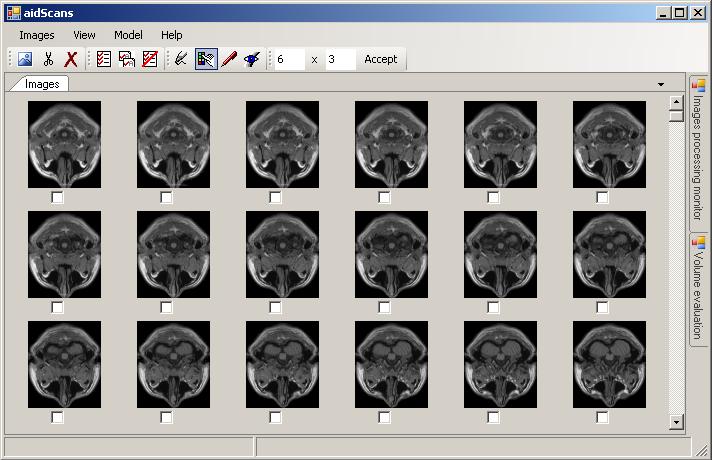
The aidScans is able to render 3D-models of objects. The head model you can see below was reconstructed using set of MRI slices. The model itself, points and outlines, was obtained by the aidReady library.
170 head slices. Ones were received from a MRI tomograph.
The head model was reconstructed using slices listed before.
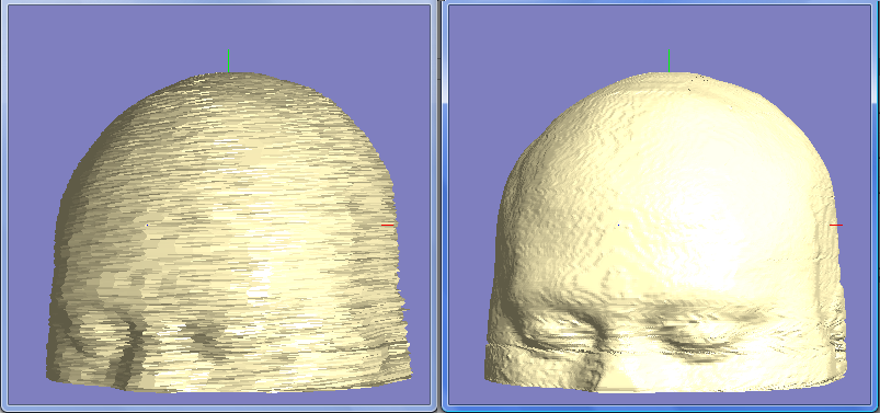
The head model was reconstructed using slices listed before.
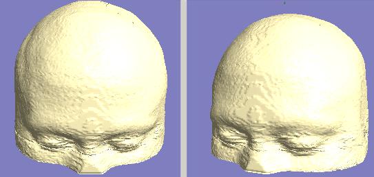
The head model was reconstructed using slices listed before.

The head model was reconstructed using slices listed before.
